i-CAT 3D CT Scan in Tuckerton, NJ for Advanced Dental Imaging and Precise Care
 At Tuckerton Dental, we are dedicated to providing patients with the latest dental technologies for accurate diagnosis and treatment. One of the most advanced tools we offer is the i-CAT 3D CT scan. This technology allows Dr. Ronald Petrosky, a leading dentist in Tuckerton, NJ, to see detailed images of your teeth, jawbone, and surrounding structures in three dimensions. Patients in Tuckerton, Little Egg Harbor, Bass River, Beach Haven, and nearby areas can benefit from this precise imaging, which helps ensure that every procedure is planned and executed with accuracy.
Whether you are considering dental implants, orthodontics, or other complex procedures, i-CAT 3D imaging near you provides the insight necessary for safe, effective care. Dr. Petrosky’s extensive training in this technology ensures that patients receive the highest quality of treatment available. To learn more about the purpose and benefits of 3D imaging, contact our Tuckerton dental office by calling (609) 296-1007.
Why Tuckerton Dental is the Best Choice for 3D Imaging
At Tuckerton Dental, Dr. Ronald Petrosky combines his advanced training with years of clinical experience. He has completed 12 hours of operating training on the i-CAT Cone Beam 3D Dental Imaging System and the professional course Implementation and Utilization of Cone Beam 3D Imaging in Your Practice. This specialized training allows him to operate the technology with precision and use the results to guide treatment decisions effectively.
By choosing Dr. Petrosky, you can feel confident that your dental care is guided by a dentist who combines years of experience with advanced technological expertise. He has helped countless patients in Tuckerton, Little Egg Harbor, Bass River, Beach Haven, and the surrounding areas achieve successful outcomes with the assistance of 3D imaging.
What is an i-CAT 3D CT Scan?
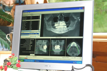
An i-CAT 3D Cone Beam CT scan is a type of dental imaging that creates a comprehensive three-dimensional view of your mouth, teeth, jaws, nerve pathways, and sinuses. Unlike traditional X-rays, which show only flat images, 3D imaging provides a complete picture of your oral structures.
This technology is particularly useful for patients who require implants, orthodontic adjustments, or evaluation of complex dental conditions. The i-CAT 3D scan captures detailed information that can help identify hidden issues, improve treatment planning, and increase the success of your dental procedures.
Benefits of i-CAT 3D Imaging
 Precision for Better Treatment Planning
 Precision for Better Treatment Planning
The i-CAT offers the highest level of surgical predictability, resulting in more successful outcomes for patients. Detailed 3D images allow Dr. Petrosky to identify potential issues and plan dental procedures with precision.
Safety and Low Radiation
i-CAT 3D scans use over ten times less radiation than a traditional medical CT, making them a safer option for most patients.
Fast, Efficient, and Accurate
A quick 8.9-second scan produces the most anatomically accurate 3D images of the mouth, face, and jaw. Within minutes, Dr. Petrosky can offer an immediate virtual diagnosis and treatment plan, all in one visit.
Reduced Surgery Time and Complications
By clearly visualizing vital structures such as nerves and sinuses, i-CAT scans help reduce surgery time and minimize the risk of complications.
Comfort and Convenience
The i-CAT’s open-environment design increases patient comfort, and the scan is non-invasive and painless. Patients can avoid hospital visits and multiple appointments, saving time and stress.
Cost-Effective and Collaborative
i-CAT imaging is more affordable than a medical CT scan and allows Dr. Petrosky to easily share the 3D data with a patient’s orthodontist, oral surgeon, or other medical practitioners using free i-CAT Vision Software, ensuring coordinated care.
Common Uses for i-CAT 3D Imaging
The i-CAT 3D scan is versatile and can be used in many areas of dental care. Patients who may benefit include those needing:
- Dental implants: The scan provides precise measurements for safe and accurate implant placement.
- Orthodontics: Helps in planning tooth movement and evaluating jaw structure for braces or Invisalign.
- TMJ evaluations: Identifies jaw joint problems and helps guide treatment.
- Complex extractions: Assists in planning for wisdom teeth or other surgical procedures.
- Sinus or jaw issues: Detects abnormalities and allows for careful planning of corrective treatment.
Patients from Tuckerton and nearby communities such as Little Egg Harbor, Bass River, Beach Haven, and Long Beach Island often come to Tuckerton Dental for advanced imaging and personalized care.
Frequently Asked Questions
Yes. The i-CAT scan uses low-dose radiation and is considered safe for most patients, including adults and teens. It exposes you to over ten times less radiation than a traditional medical CT, making it a safer choice for dental imaging.
A typical scan takes less than one minute, specifically around 8.9 seconds. The images are digital and available immediately, allowing Dr. Petrosky to review your results and begin planning your treatment without delay.
No. The procedure is completely non-invasive and painless. You simply sit comfortably while the machine rotates around your head, capturing detailed 3D images of your teeth, jaw, and facial structures.
The highly detailed 3D images allow Dr. Petrosky to see structures that may not be visible with traditional X-rays, such as nerves, sinuses, and bone density. This helps with accurate treatment planning for implants, orthodontics, TMJ therapy, or oral surgery, reducing risks and increasing the likelihood of a successful outcome.
Experience Advanced Imaging for Confident Dental Care at Tuckerton Dental
The i-CAT 3D CT scan is an essential tool for precise, safe, and effective dental care. At Tuckerton Dental, Dr. Ronald Petrosky uses this technology to provide patients in Tuckerton, NJ, and the surrounding areas with a higher level of care. Whether you are considering dental implants, orthodontics, or other complex procedures, the detailed imaging allows your dentist to plan every step with confidence.
Schedule your appointment today with the top dentist in Tuckerton, NJ by calling (609) 296-1007 to experience the benefits of advanced 3D imaging for your dental care.